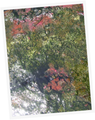
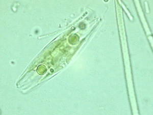
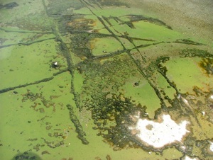
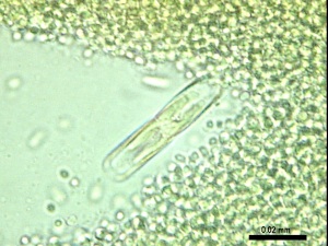
Magadi Hot Springs

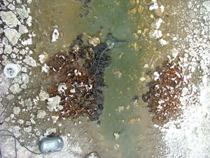
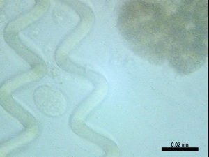
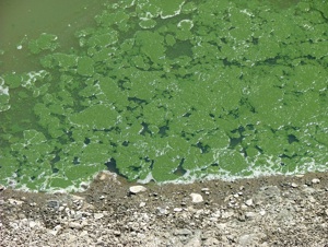
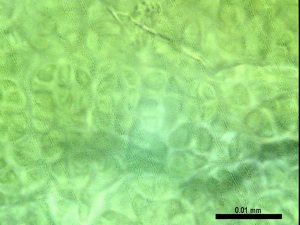
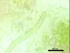
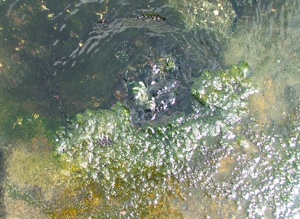
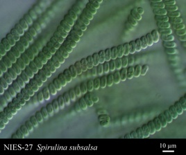
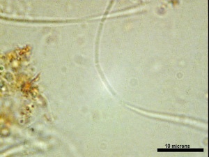
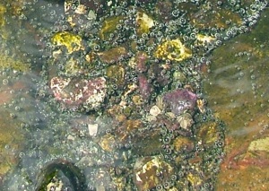
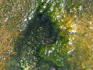
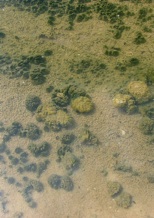
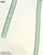
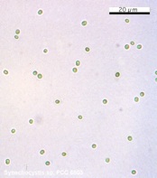
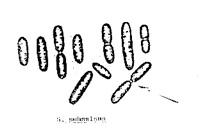
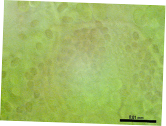
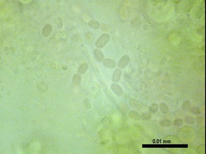
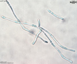
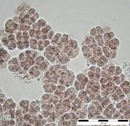
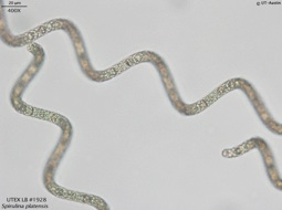
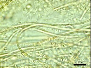
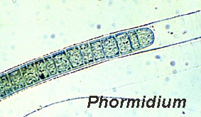
One of the tourist draws of the Magadi area is the number of hot springs that feed the lake. Living in these springs is a number of bacteria and some algae, and here I would like to briefly categorize the different types you might find out there. Essentially I tried to sample as many distinctive forms around the hot spring as possible, but, being a Geologist, I’m sure a biologically oriented person would be able to find many more then I did, especially if more sophisticated molecular methods were employed. So, think of this as a brief introduction to the things in the springs, and realize there are probably many more waiting to make your acquaintance.

Things in the Springs

These ridges are pretty striking when you are out at the hotsprings. They are about 1 inch high at the crest of the ridge, are submerged in shallow water and as can be seen in the above photo, they are very linear and growth is pretty much restricted to right around the ridge (the dark coloration is where growth is). The ridges are in standing water away from the vigorous springs, and the growth around them are green-brown in color. My guess is that these represent growth around fractures where brines can seep up and bring nutrients to the bacteria. Under the microscope, this sample seems to be dominated by green filaments in large, fibrous, hay-like clumps. These filaments are segmented under close examination (right side of image below sample location) and it is believed that the bulk of this sample is Oscillatoria as seen above right. There are also sheets of small spheres and occasional diatoms (large cell to the left of imaged filaments) in the sample.

A1: Ridge Growths

Temp: 41.5 °C

Na+: 7900 ppm

pH: 10.2

K+: 98 ppm

Tentative Identification: Oscillatoria


This sample was found at the surface of the water, was yellow-green, slimy in texture, and trapped large bubbles in a film (indicated in the top picture by a yellow arrow). In this picture the sampled growth is at the water’s surface right above a spring outflow (you can see a arced distortion in the water’s surface above the non-pointer end of the arrow which is from the force of the flowing water from the spring).  This sample is dominated by small green spheres, with occasional filaments, corkscrews and, as pictured above, diatoms. It is believed that the green spheres that dominate the sample are Synechocystis (or Microcystis) cells. Microcystis is known to produce buoyant colonies due to the presence of gas vesicles in the cells, but these vesicles are likely not resolved in these photos.

A2: Surface algae

Temp: 42.8 °C

Na+: 7500 ppm

pH: 10.2

K+: 86 ppm

Tentative Identification: Synechocystis

This red mat was found right at the mouth of a small spring, and was almost covered by green growth. In 2005, when it was wetter, the red patches were much larger in area. I have a feeling that the distribution of the different spring critters changes during the seasons as water depth, or salt concentration changes.


Under the microscope this sample was seen to be composed of fine filaments which have segments 3-5 microns long enclosed in a sheath that often runs past the terminal cell. Also in this sample were small green spherical cells similar to those seen in A2. This sample was identified as Leptolyngbya, which is a mat forming genus typified by narrow cell width. This genus includes organisms that would have previously been classified as Phormidium. It should be noted that since the bulk of the sample was cyanobacteria, it may be possible that actual sampling of the red mat failed. The amount of red material available to sample in the field was very small, so a neighboring growth may have been identified instead.

A3: Red Growth (?)

Temp: 41.8° C

Na+: 7500 ppm

pH: 10.3

K+: 86 ppm

Tentative Identification:Leptolyngbya

This was purple crust was found covering a small pebble. This sample had some fibrous red cells like in A3 above, but is mostly dominated by spherical/oblong green and red cells. Some of these oblong cells may be Synechococcus, however, identification was difficult. Additionally, some Spirulina was found in dense clumps in sections of the sample. As with A3, it may be possible that the organisms responsible for the purple color were not actually examined.

A4: Purple Algae (?)

Temp: 43.4 °C

Na+: 7500 ppm

pH: 10.3

K+: 82 ppm

Tentative Identification: Synechococcus and Spirulina

In the field, these were growing as a dark green mats/pillars right near a hotspring outlet. They had a gritty texture and seemed to be the only major growth occupying this area. The same variety seemed to be in other, mostly bare areas with fine gritty substrate near or in hotspring outlets. The sample collected was found in the hottest water sampled in this collection effort.

This was a hard sample to examine due to all the detrital material caught up with the biologic sample (it made the cover slip not want to lay flat). The overall red color might be from inorganic (iron?) material, and as for the biology there seemed to be several layers (which also made viewing difficult). There were green ovoid cells, but the bulk of the sample was composed of segmented filaments as seen above. These are thought to be Leptolyngbya  (possibly would have originally been called Schizothrix, but is not treated as a distinct genus in Bergey’s manuals).

A5: Dark green pillars

Temp: 44.4°C

Na+: 7900 ppm

pH: 10.2

K+: 90 ppm

Tentative Identification: Leptolyngbya (Schizothrix?)

This stuff can be found growing all around the weaker springs and seems to have little or no competition from other types of visible growths. It has a distinctive red-brown color, but it was noticed that my sample may have lost the red-brown pigments while in storage. The tips of the preserved sample were white and colorless under the scope, while the rest was green. Most of the cells are are green spheres, and there are a few rods concentrated in hair-like masses. It is thought that this sample may be colonies of Pleurocapsa or Myxosarcina. Some baeocytes were seen in this sample, with the number/parent cell being more indicative of Pleurocapsa, but the overall growth form seems more like Myxosarcina... but Pleurocapsa can have a similar form in certain growth conditions.

A6: Red-Brown mats

Temp: 34.8 ° C

Na+: 8250 ppm

pH: 10.4

K+: 100 ppm

Tentative Identification: Pleurocapsa/ Myxosarcina

Near the salt ponds there was a fresher pond that was full of these green floating growths, and the water itself was also green in color. Under the scope the vast majority of the cells were featureless green spirals but there were small clumps of spheres (such as the one between the two spirals imaged from the sample) in addition to a few spirals that were composed of spherical segments (the upper right part of the magnified sample is thought to be a coiled Anabaenopsis). The loosely spiraled cells that compose the bulk of the sample is Arthrospira (often referred to as Spirulina, but they are considered distinct genera at present). This pond in particular hosted a number of different bird species, including flamingos, which are known to feed on Arthrospira.

A7: Green Lagoon

Temp: NA

Na+: 5100 ppm

pH:  ~11

K+: 315 ppm

Tentative Identification: Arthrospira

Image Source: NIES