Published in The Picking Table 43(2),
11-13 (2002).
GROWTH SPIRALS ON
GRAPHITE CRYSTALS FROM THE
TROTTER MINE DUMP, FRANKLIN, NEW JERSEY
John A. Jaszczak
Department of Physics and the A. E. Seaman Mineral
Museum
Michigan Technological University
1400 Townsend Dr.
Houghton, Michigan
49931-1295
John Rakovan
Department of Geology
Miami University
Oxford, Ohio
45056
The Franklin and Sterling Hill deposits have been the source of several mineralogically important graphite occurrences. While the Franklin Marble is rich in "ordinary," small, disseminated graphite flakes, several notable occurrences of graphite have been found in and near the Franklin and Sterling Hill deposits. Some examples include the remarkable barrel-shaped crystals (the best ones not exceeding 0.5 mm) described by Charles Palache from the 900' level of the Sterling Mine in 1941 (Palache, 1941; Jaszczak, 1994a, 2001), and the spherical graphite aggregates, ranging from sub-millimeter to 2 cm across, that have been found at both deposits (Lemanski, 1991; Jaszczak, 1994b, 1995; Hanna and Jaszczak, 1999).
New to the list of remarkable graphite crystals from this area are crystals up to 2 mm across that show visible growth spirals on the basal pinacoid faces c{001}. While graphite crystals are common throughout much of the world, those showing pronounced growth spirals are relatively rare. Such crystals have been noted on only a small fraction of the crystals occurring at several localities, which are listed in Table 1.
TABLE
1. Occurrences of growth spirals on natural graphite crystals.
|
Locality |
Reference |
|
Canada:
Princeton Resources Corp. mine, Bissett Creek, Renfrew County, Ontario. |
Jaszczak (unpublished). |
|
Namibia:
road D-1918 heading for Big Spitzkoppe, near Usakos. |
Jaszczak (unpublished). |
|
Namibia:
near Wlotzkas Baken. |
Weiner and Hager, 1987; McCall et al.
1999, Rakovan and Jaszczak, 2002 |
|
Ukraine:
Zavallya deposit in Bug River area; Smela anorthosite complex, Ukrainian
Shield; Stary Krym deposit, Azov Sea area. |
Kvasnitsa and Yatsenko, 1997; Kvasnitsa et al., 1999. |
|
U.S.A.:
Lime Crest quarry, Sparta, New Jersey. |
Jaszczak, 1998. |
|
U.S.A.:
Jensen quarry, Riverside County, California. |
Jaszczak (unpublished). |
|
U.S.A.:
Crestmore quarry, California. |
Jaszczak, 1991. |
|
U.S.A.:
Gouverneur Talc Company No. 4. quarry (Valentine Deposit), Harrisville, New
York. |
Chamberlain et
al., 1999. |
|
U.S.A.:
Trotter Mine dump, Franklin, New Jersey. |
This work. |
|
U.S.A.
and Canada: Grenville series limestones. |
Horn, 1952. |
During the April 24-25, 1999 collecting trip to the Trotter mine dump, Franklin, New Jersey, Mr. Wayne Cokeley collected samples of graphite crystals in marble from a series of large boulders near the edge of the dump. The graphite occurs in these samples as disseminated, tabular flakes as much as 4 mm across associated with abundant, amber phlogopite crystals in calcite. Tiny, spheroidal graphite aggregates occur on some of the phlogopite crystals. The calcite matrix is tan-colored moderately fluoresces orange-red in short-wave ultraviolet light, which indicates that the samples probably originated from within the orebody or within its manganese halo (Richard Bostwick, personal communication, 2000). The fluorescence is also in contrast to otherwise similar-looking material from the Lime Crest quarry, Sparta, New Jersey (Jaszczak, 1998) which typically does not fluoresce. Etching of the marble in dilute hydrochloric acid revealed the presence of some light-colored insoluble crystalline masses that resemble pyroxenes or amphiboles but were not positively identified.
The growth spirals on the graphite crystals are easily visible in reflected light with a stereoscopic microscope. On the crystal in Fig. 1a, the steps are fairly straight and uniformly separated from one another. The corners of the steps and the steps near the center of the spiral are rounded. The step edges appear to be more or less parallel to <100>, the outline of the crystals formed by the edges between the basal pinacoid and the dominant prism (probably first-order) faces. A second crystal (Fig. 1b) situated less than 5 mm from that in Fig. 1a, shows less regularity in the step spacing, although the step edges are also more or less parallel to the edges of the external crystal form. This crystal also shows some surface pitting and some wavy step edges, particularly near the center of the crystal, which may indicate that some degree of dissolution took place. On a surface of the host rock approximately 30 cm2, approximately 35 graphite crystals were exposed; however, only the two illustrated here show visible growth spirals.
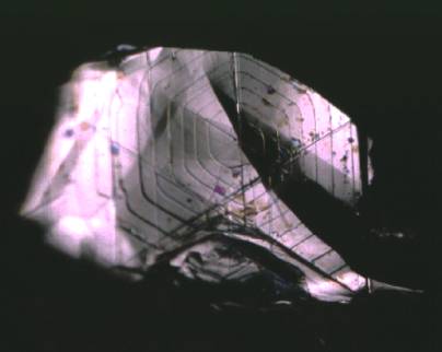
(a)
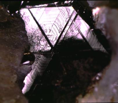
(b)
Fig. 1. Growth spirals on tabular graphite crystals in calcite from the Trotter Mine dump, Franklin, New Jersey. Both crystals are 2 mm across and occurred within 5 mm of each other. (J.A. Jaszczak specimen #2336 and photos 83-29, 83-24.)
The occurrence of prominent growth spirals on the surface of the graphite crystals may give some clues as to their origin. Such spirals are indicative of graphite growth by the spiral growth mechanism (Sunagawa, 1987) first proposed by Frank (1949). Spiral growth is well documented in minerals that have grown from solutions and vapors, and indicates growth at relatively low supersaturations with a regular addition of growth units (atoms or clusters of atoms) to specific surface sites, especially steps. Graphite disseminated in metasedimentary rocks is generally believed to be formed from the graphitization of organic matter in precursor sediments during metamorphism (see, for example: Weis et al., 1981; Buseck and Huang, 1985; Wopenka and Pasteris, 1993; Luque et al., 1998). During heating, organic precursors release oxygen, hydrogen, and nitrogen, and the remaining carbon progressively orders into crystalline graphite. However, since the spiral growth mechanism is one that is mediated by dynamics at a crystal's surface, it is an extremely unlikely mechanism for growth in solid-state transformations such as the graphitization of carbonaceous matter where crystallization takes place throughout the bulk of the developing crystal. It seems more likely that the crystals grew by carbon precipitation from a carbon-rich fluid permeating the system (see, for example: Rumble and Hoering 1986; Luque et al. 1998; Satish-Kumar 2001), perhaps after peak metamorphism, but at relatively low supersaturation and unhindered by surrounding calcite. Although graphite is usually considered a fairly chemically inert mineral, it is well known that it can be dissolved [see for example Jaszczak (1991) Fig. 12] or precipitated from high temperature and pressure carbon-oxygen-hydrogen fluids. The carbon could have originated from organic matter in the sediments [this would be indicated by relatively light carbon isotopic ratios, 13C/12C, for the graphite], and mobilized under later fluid flow (Crawford and Valley, 1990). Graphite could have subsequently re-precipitated under changing conditions, such as regional cooling or changing solution composition due to fluid-rock reactions. Although the picture is incomplete, growth spirals on the surface of graphite crystals are an attractive piece in the puzzle of understanding their genesis.
ACKNOWLEDGMENTS
We are grateful to Michael P. Basal and Wayne Cokeley for supplying material from which these crystals were discovered. Many thanks are also due to Steve Phillips for allowing collectors access to the site, and to Don Halterman of the Delaware Valley Earth Science Society for collaborating with Mr. Phillips to make the 1999 collecting event possible.
REFERENCES
Buseck, P. R. and Huang,
B.-J. (1985) Conversion of carbonaceous material to graphite during
metamorphism. Geochimica et Cosmochimica
Acta 49, 2003-2016.
Chamberlain, S. C., King, V.
T., Cooke, D.; Robinson, G. W. and Holt, W. (1999) Minerals of the Gouverneur
Talc Company No. 4 quarry (Valentine Deposit), Town of Diana, Lewis County, New
York Rocks & Minerals 74, 236-249.
Crawford, W. A. and Valley, J. W. (1990) Origin of graphite
in the Pickering gneiss and the Franklin marble, Honey Brook Upland,
Pennsylvania Piedmont. Geological Society
of America Bulletin 102,
807-811.
Frank,
F. C. (1949) The influence of dislocations on crystal growth. Discussions of the Faraday Society 5, 48-54.
Hanna,
G. A. and Jaszczak, J. A. (1999) A new find of spherical graphite from Sterling
Hill, New Jersey. The Picking Table 40, 27-30.
Horn,
F. H. (1952) Spiral growth on graphite. Nature
(London) 170, 581.
Jaszczak, J. A. (1991)
Graphite from Crestmore, California. Mineralogical
Record 22, 427-32.
Jaszczak,
J. A. (1994a) Famous graphite crystals from Sterling Hill, New Jersey. The Picking Table 35(2), 6-11.
Jaszczak, J. A. (1994b) On
the Natural Occurrence of Spherical Graphite. Rocks & Minerals 69,
117-118.
Jaszczak, J. A. (1995)
Graphite: Flat, Fibrous and Spherical.
In Mesomolecules: From Molecules
to Materials, ed. by G. D. Mendenhall, J. Liebman, and A. Greenberg
(Chapman & Hall, New York) pp. 161-80.
Jaszczak, J. A. (1998)
Unusual graphite crystals from the Lime Crest quarry, Sparta, New Jersey. The Picking Table, 39(1), 20-24; and (1997) Rocks
& Minerals 72, 330-334.
Jaszczak,
J. A. (2001) Palache's "Contributions to the Mineralogy of Mineralogy of Sterling
Hill, New Jersey": the 900-foot level revisited. The Picking Table 42(1),
6-15.
Kvasnitsa, V. N. and
Yatsenko, V. G. (1997) Growth spirals on graphite crystals from Ukraine. Mineralogicheskii Zhurnal 19(6), 43-48.
Kvasnitsa, V. N., Yatsenko, V.
G. and Jaszczak, J. A. (1999) Disclinations in unusual graphite crystals from
anorthosites of Ukraine. Canadian
Mineralogist 35, 951-960.
Lemanski, C. S., Jr. (1991)
Graphite in ore: An unusual occurrence at the Sterling mine. The Picking Table, 32(1), 11-3.
Luque, F. J.; Pasteris, J.
D.; Wopenka, B.; Rodas, M.; and Barrenchea, J. F. (1999) Natural
fluid-deposited graphite: Mineralogical characteristics and mechanisms of
formation. American Journal of Science 298,
471-498.
McCall, K., Rakovan, J., and
Jaszczak, J. (1999) Multiple length scale growth spirals on natural graphite
(001) surfaces. GSA Abstracts with
Programs 31(7), A-169.
Palache, C. (1941)
Contributions to the Mineralogy of Sterling Hill, New Jersey: Morphology of
Graphite, Arsenopyrite, Pyrite, and Arsenic. American Mineralogist 26, 709-717.
Palache, C., Berman, H., and
Frondel, C. (1944) The System of
Mineralogy of James Dwight Dana and Edward Salisbury Dana, Yale University
1837-1892. Seventh edition, vol. 1. (John Wiley and Sons, New York).
Rakovan, J. and Jaszczak, J. A.
(2002) Multiple length scale growth spirals on metamorphic graphite {001}
surfaces studied by atomic force microscopy. American Mineralogist 87,
17-24.
Rumble, D. III, and Hoering, T.
C. (1986) Carbon isotope geochemistry of graphite vein deposits from New
Hampshire, U.S.A. Geochimica et
Cosmochimica Acta 50, 1239-1247.
Satish-Kumar, M., Wada, H.,
Santosh, M. and Yoshida, M. (2001) Fluid-rock history of granulite facies
humite-marbles from Ambasamudram, southern India. Journal of Metamorphic Geology 19,
395-410.
Sunagawa, I. (1987) Surface
Microtopography of Crystal Faces. In Morphology
of Crystals. I Sunagawa, editor. (Terra Scientific Publishing Company,
Tokyo) pp. 321-365.
Weiner, K.-L. and Hager, H.
(1987) [Growth spirals on graphite crystals.] Lapis 12(1), 31-33. [In
German].
Weis, P. L., Friedman, I.,
and Gleason, J. P. (1981) The origin of epigenetic graphite: evidence from
isotopes. Geochimica et Cosmochimica Acta
45, 2325-2332.
Wopenka, B. and Pasteris, J. D. (1993) Structural characterization of kerogens to granulite-facies graphite: Applicability of Raman microprobe spectroscopy. American Mineralogist 78, 533-577.