It is clinically important to know the oxygen saturation of the blood under ambulatory conditions. Low amount of oxygen in the arterial system would result in onset of hypoxia, and could lead to tissue damage. Therefore, a system which would detect the presence of blood circulation as well as the amount of oxygen in the blood is needed. Pulse oximeters are the most commonly used tool for this particular purpose.
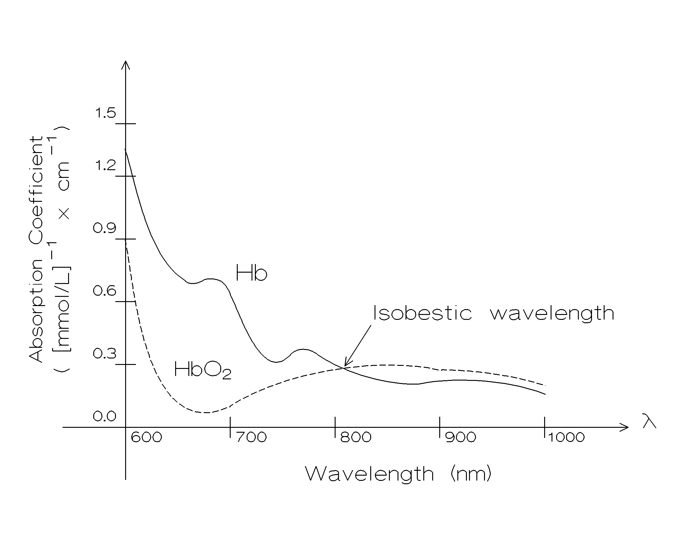
Hemoglobin is the protein contained in the red blood cells which help carry the oxygen in the blood. 98 % of the oxygen carried by the blood is carried by the hemoglobin while the remaining 2 % is dissolved in the plasma portion of the blood. Pulse oximeters take advantage of the fact that the hemoglobin has different light absorption characteristics at different wavelengths. Optical absorption characteristics of oxygenated and de-oxygenated hemoglobin are shown on Figure 1 below. As it can be seen from the traces, the two spectra intersect at the isobestic wavelength of l =805 nm, where the absortion is independent of oxygen saturation in the blood. Absorption measured at this frequency is used as a reference value. The two spectra differ greatly at l =660 nm, which used as the measurement point. Commercial unit that is available in the laboratory uses l =660 nm measurement and l =910 nm for reference where the choices are dictated usually be the availability of components and competitive patent coverage.
Figure 1. Optical absorption spectra of oxygenated (HbO2) and de-oxygenated hemoglobin (Hb).
Absorption of light as it passes through the tissue can be measured by shining a light onto the tissue and measuring the received light on the opposite site. If the wavelength of the incident light is known, then the absorption can be determined as
![]() , [ Eq. 1 ]
, [ Eq. 1 ]
where
IR is the intensity of the received light,IT is the intensity of the transmitted light,
a is the absorption coefficient,
c is the concentration of the absorbing molecule (Hb or HBO2)
d is the thickness of the tissue.
The term a.c.d is known as the optical density of the tissue, and expressed as
![]() [ Eq. 2 ]
[ Eq. 2 ]
In the case of pulse oximetery, the optical density is determined at two wavelengths and the Sa(O2) is determined using the following relationship:
![]() [ Eq. 3 ]
[ Eq. 3 ]
where the coefficients A and B must be determined experimentally.
Above described system is also able to detect the heart beats by sensing the changes in the blood volume following systole. As more blood enters the tissue after each beat, absorption characteristics of the tissue changes, which is reflected in the IR.
EXPERIMENT:
In this experiment we will use a portable pulse oximeter, Nonin Model 9843.
REPORT:
|
Signal 1 Strength |
Signal 1 Strength |
|
|
Empty |
||
|
Finger |
||
|
Pink Filter |
||
|
Green Filter |