Although the respiratory system is very complex and its analysis requires identification of multitude of parameters, in practice only very few of them can be measured, especially if the methods are required to be non-invasive. These parameters include the volume of the air flowing through the mouth and nose, partial pressures of the gasses passing through the airways and the temperature of the air. However, it is surprising that many other parameters of the respiratory system can be estimated from the measured quantities.
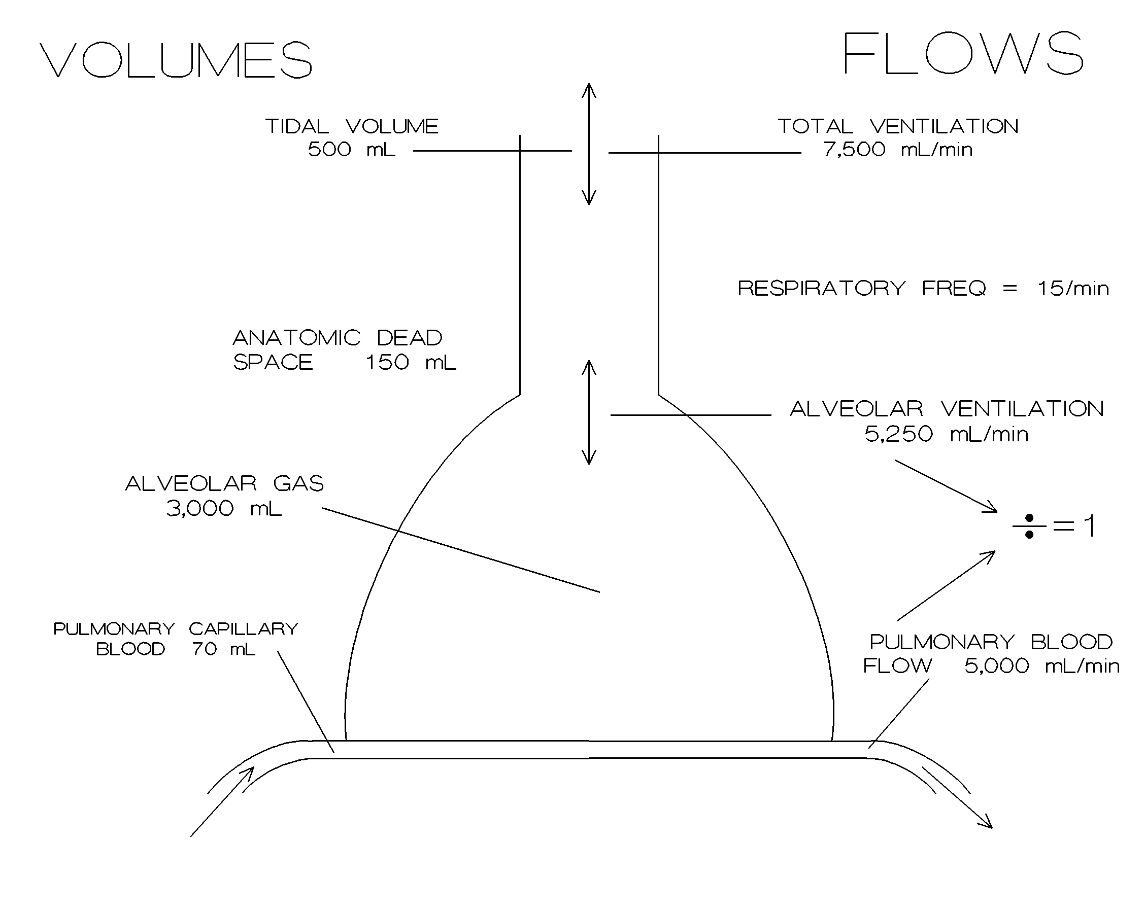
Figure 1. Diagram of lung showing typical volumes and air flow.
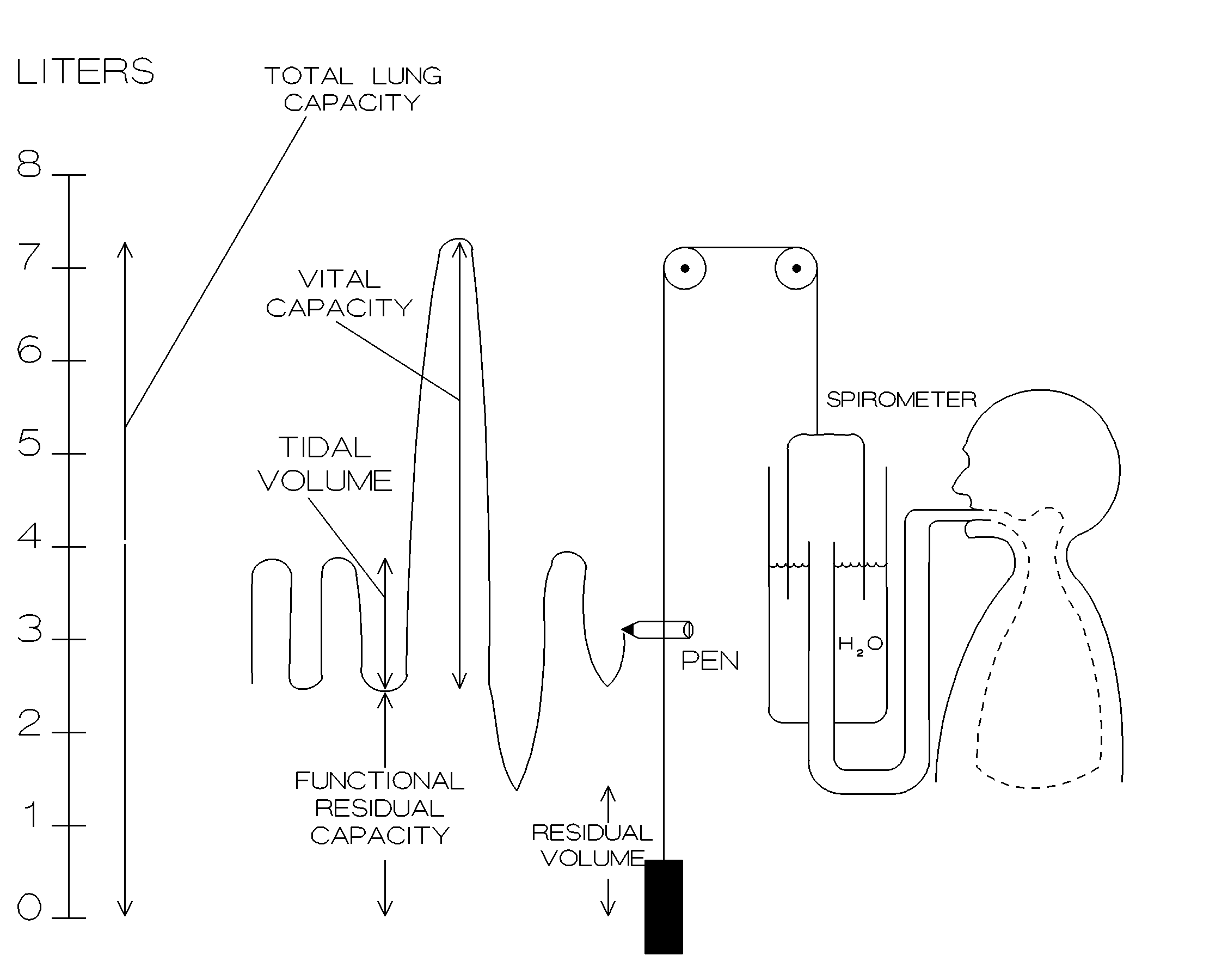
Figure 2. Basic spirometry and measured lung volumes.
Figure 1 above shows the volumes and the flow rates of air at the various parts of the lungs. Figure 2 shows the principle of operation of a spirometer as well as the definitions of various measured lung volumes. Spirometer measures the direct air volume and allows the measurement of all volumetric parameters except the residual volume.
Another way to measure the volume of air inhaled or exhaled is through the measurement of flow of air. Gas flow rates can be measured by rotating vane flow meters, ultrasonic flowmeters, thermal convection flowmeters, or by differential pressure flowmeters. Following paragraphs should provide a brief discussion of each technique.
Rotating vane flow meters utilize a small turbine in the flow path of air. Rotation of the turbine can be related to the air flow and the exchanged air volume. Optical, electronic and mechanical detection systems are all used for this purpose.
Ultrasonic transducers utilize the changes in the frequency of the received signal or the changes in the transit time to estimate the flow velocity, as it was explained in the write up for experiments in Week 5. Reader should review those techniques for further information.
Thermal convection flow meters (also known as the nasal thermistors) utilize the changes in the temperature to estimate the air flow. If the average temperature of the sensor is kept at a value somewhere between the ambient temperature and the body temperature, then any transient drop in the temperature would be due to flow of cold room air over the thermistor. On the other hand, any sudden rise in the temperature would be due to exhaled warm air from the lungs. Principle of operation of this type of flow sensor is very similar to the thermo-dilution method of flow measurements as it was discussed in the write up of week 5, and it could be reviewed for further information.
Differential pressure flowmeters (also known as pneumotachometers) consist of two pressure transducers placed at opposite ends of a restriction tube with known resistance to flow. Flow than can be calculated as the ratio between the pressure differential and the tube's resistance to flow as shown below:
![]() [ Eq. 1 ]
[ Eq. 1 ]
where f is the flow, P1 and P2 are the pressure values measured at the ends of the restriction tube, and R is the tube's resistance to flow.
For all methods discussed above for the measurement of the flow, one needs to calculate the time integral of the flow to find the changes in the lung volume,
![]() [ Eq. 2 ]
[ Eq. 2 ]
Although the use of methods discussed above usually allow accurate estimates of air flow and lung volume changes, they are somewhat invasive and all of them require the cooperation and diligence of the patient. For non-conscious or sleeping adults, or infants and small children, a method where the subject does not have to keep the flow transducer in their mouth is needed. Inductance plethysmography is such a method.
BACKGROUND ON INDUCTANCE PLETHYSMOGRAPHY
In inductance plethysmography, subject wears two elastic bands, one around the chest, other around the abdominal area as shown in Figure 3 below. A conductor in each one of these elastic bands form an inductor. An oscillator sends sinusoidal signals to the inductor and measures the changes in the cross sectional area of the inductor. Using this information, one can deduce the area enclosed by the inductor, hence the thoracic and abdominal cross sectional area.
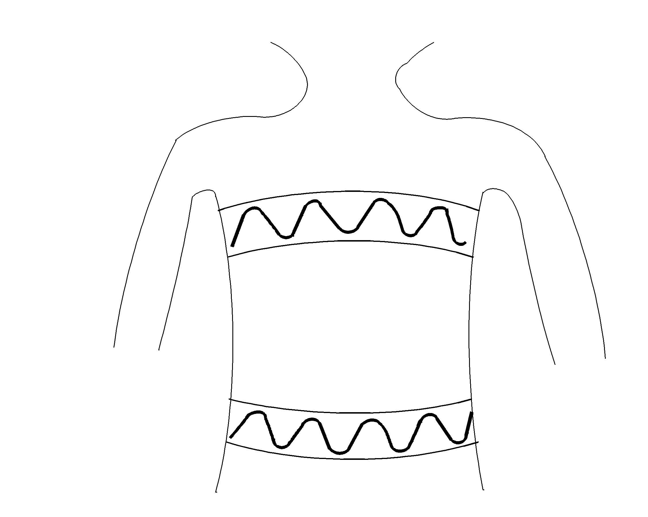
Figure 3. Basic inductance plethysmography setup.
Inductance of any conductor is given by
 [ Eq. 3 ]
Voltage induced on the conductor can be determined using the Faraday's Law of Induction:
![]() [ Eq. 4 ]
[ Eq. 4 ]
where the magnetic field B(t) is also produced by the current I(t).
Ampere's Law can be used to calculate the magnetic field, B(t), if the entire structure is symmetric. In a circular loop, symmetry holds true only for the center of the loop, but not for any other point within the loop. Therefore, we must use the Biot-Savart Law to calculate the magnetic field within the loop:
![]() [ Eq. 5 ]
[ Eq. 5 ]
where m 0=4p x10-7,
I is the current on the wire segment ![]()
r is the distance between the wire segment and the point where the magnetic field is being measured.
One can integrate equation 5 along the wire loop forming the coil to find B(t), which can be plugged into the equation 4 to determine the voltage. Finally the inductance can be found using the equation 3. Even for a simple shape like a circle, the above equations are difficult to calculate. Engineers usually utilize numerical techniques to calculate such integrals. Figure 4 below shows the results from numerical evaluation of inductor values as a function of the radius of the circular loop. As it can be seen from the figure below, inductance increases linearly with the radius, or by the square root of the area of the circle.
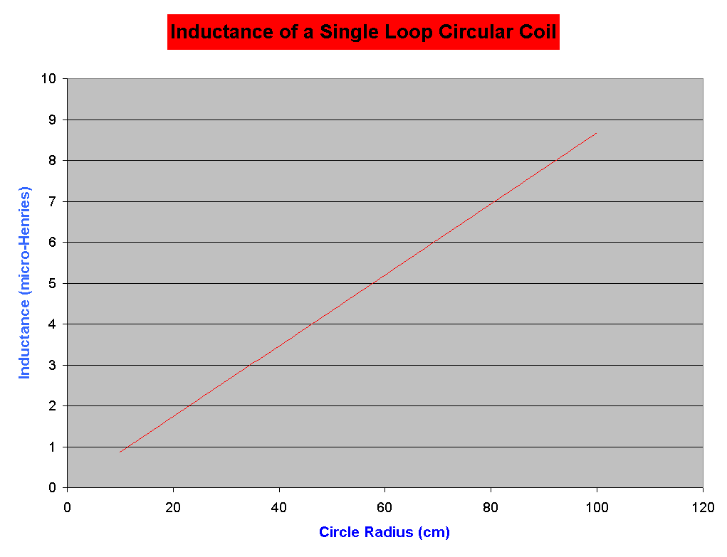 Figure 4. Inductance values calculated from numerical simulations.
Inductance plethysmograph instruments measure the inductance of the two coils as seen in Figure 3. From this measurement, instrument estimates the cross sectional area of the chest and the abdominal coil. Using these two calculated areas, device estimates the tidal volume during normal breathing. Reader must note that this system can only give relative measurements since there is no reliable way to relate the cross sectional area measurements to absolute respiratory volumes. If an absolute measurement is desired, then a device must be calibrated once for each patient using a spirometer.
EXPERIMENT:
REPORT: